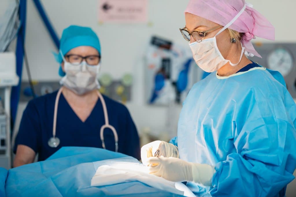
Hookwire Guided Excision of the Breast
Abnormalities can often be picked up on breast ultrasound or mammogram long before a surgeon can feel a lump or a woman has any symptoms. This means that Dr Adams needs a guide to the location of the tumour so she can remove the abnormal area from the breast.
A hookwire is a thin wire placed in the breast which helps Dr Adams surgically remove breast abnormalities she cannot feel. They can be used for either small invasive breast cancers and areas of DCIS; or other abnormalities which require a surgical biopsy where the radiological biopsy is insufficient. On the day of surgery a patient visits radiology to have the hookwire placed. The radiologist locates the abnormality with mammogram and/or ultrasound.

Local anaesthetic is used to numb the skin, and a thin flexible wire not much thicker than fishing line is placed alongside the abnormal area in the breast. Repeat ultrasound and mammogram are then taken. The external portion is then taped down flush to the skin and the patient is transferred to the operating room. During the operation Dr Adams uses the wire as a guide to excise the correct area of the breast. Whilst the patient is still asleep, imaging of the excised tissue is usually performed to confirm the abnormality has been excised completely. The sample is then sent away for pathological assessment to confirm the nature of the lesion for excisional biopsy and for more detailed assessment of surgical margins for cancerous tumours.
The surgery is done under general anaesthetic and usually takes between one to one and a half hours. It can be done either as a day surgery or with a single night stay in hospital. Most women recover quite quickly from surgery. There may be pain but usually this is not severe, and pain relief will be given to you prior to discharge from hospital. Bruising and tenderness of the breast is common.
It is impossible to perform this surgery and not affect the appearance of the breast. Scars are unavoidable with all surgery. Where possible they are placed at the edge of the areola though this depends on the location of the abnormality. Dr Adams places great emphasis on the appearance of the breast after surgery but some changes can occur. In addition to a scar there can be a change in the shape or size of the breast. This is usually more common in surgery for cancer as more tissue is taken and patients often require radiotherapy. Where possible oncoplastic techniques are used to close the breast cavity and achieve the best cosmetic outcomes possible for patients.
Dr Adams will meet with you approximately one week after surgery. At this appointment a general review is undertaken and your histology or microscope report is discussed. If there was an unexpected cancer finding often more surgery is required. In cases where the surgery was performed for known cancer or DCIS, Dr Adams can discuss again what further treatment is required (see breast conserving surgery).

